
Comparison between confocal scanning laser tomography, scanning laser polarimetry and optical coherence tomography on the ability to detect localised retinal nerve fibre layer defects in glaucoma patients | British Journal of Ophthalmology

Diagnostics | Free Full-Text | Thicker Retinal Nerve Fiber Layer with Age among Schoolchildren: The Hong Kong Children Eye Study

Optical Coherence Tomography & Neurology - Practical Neurology

The Dangers of Laser Pens | Hughes, Macdonald & Davidson | Ophthalmic Opticians, with Practices in Peterhead, Inverurie, Turriff & Banchory.

Artifacts and Anatomical Variations in Optical Coherence Tomography | SpringerLink

What OCT Tells Us About Progression

figure1_38.jpg

OCT and Glaucoma: Case Review | SpringerLink

Figure 1 | British Journal of Ophthalmology

The use of Confocal Scanning Laser Tomography in the Evaluation of Progression in Glaucoma | IntechOpen

Diagnostic Imaging Devices I OCT | Heidelberg Engineering

Diagnostic Imaging Devices I OCT | Heidelberg Engineering
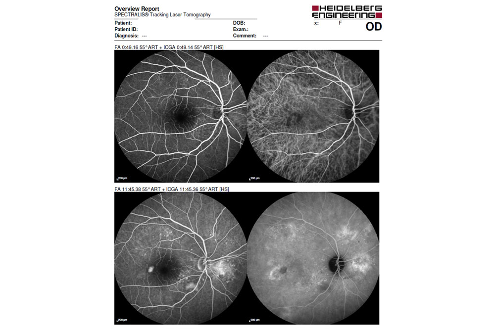
Tracking Laser Tomography. Irreversible bilateral macula edema was... | Download Scientific Diagram

An optical coherence tomography system capable of automatically and accurately imaging eyes without the use of chinrests – Duke OTC

Plasmonic Gold Nanostar-Enhanced Multimodal Photoacoustic Microscopy and Optical Coherence Tomography Molecular Imaging To Evaluate Choroidal Neovascularization | ACS Sensors

The Anatomy of an OCT Scan
; "Angiography")
Angiography

The use of Confocal Scanning Laser Tomography in the Evaluation of Progression in Glaucoma | IntechOpen

Laser Tomography - an overview | ScienceDirect Topics

3 Optical Coherence Tomography of the Optic Nerve | Ento Key
Vitreous hyper-reflective dots in pseudophakic cystoid macular edema assessed with optical coherence tomography | PLOS ONE

JCM | Free Full-Text | Optical Coherence Tomography Angiography in Diabetes and Diabetic Retinopathy

The use of Confocal Scanning Laser Tomography in the Evaluation of Progression in Glaucoma | IntechOpen

Retinal Physician - The Next Step in OCT Technology

is a SPECTRALIS printout with the upper part showing the ONH analysis,... | Download Scientific Diagram
